Join thousands of book lovers
Sign up to our newsletter and receive discounts and inspiration for your next reading experience.
By signing up, you agree to our Privacy Policy.You can, at any time, unsubscribe from our newsletters.
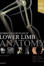
Mostly life-size dissections and osteology - corresponds to what students/practitioners will see in the dissection lab or in real life. Includes radiography and surface anatomy pictures - helps maximise clinical relevance (and necessary for modern courses). Orientational and explanatory artworks - helps the reader to position on the body. Short ccompanying text - expands on the illustrations and serves as study tool. Numbered labels - helps facilitate self-testing. Appendix containing key information on Skin, Muscles, Arteries and Nerves.
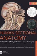
Now in its fourth edition, this portable guide and essential learning aid now contains new material. As with the previous editions, the superb full-color cadaver sections are compared with CT and MRI images. The radiological images have all been replaced with new examples for this latest edition, taken on the most up-to date equipment to ensure excellent visualization of the anatomy. The photographic material is enhanced by useful notes with details of important anatomical and radiological features.
Sign up to our newsletter and receive discounts and inspiration for your next reading experience.
By signing up, you agree to our Privacy Policy.